
Annotate contours like an expert
ART-Plan™’s Annotate is an AI-powered, CE-marked and FDA-cleared software that provides zero-click, automatic delineation of more than 200 organs at risk (OARs) and lymph nodes with the same accuracy of clinical experts in a matter of minutes.
*Note that not all models are available in all markets
* time will vary depending on the number of structures
Expert-like
The high quality of our models comes from data that have been thoroughly delineated and reviewed by experts following international contouring guidelines.
Reliable
All our models have been meticulously and continuously evaluated to ensure each organ and lymph node model is clinically acceptable.
Always better
We are continuously working to improve and add new structures to our models to ensure we remain at the forefront of the contouring practice.
OUR CT MODELS

OARs
LNs*
- Brachial Plexus (L/R)
- Brainstem
- Cerebellum
- Chiasma
- Cochlea (L/R)
- Encephalon
- Esophagus
- Eye (L/R)
- Eye Lens (L/R)
- Glottic Larynx
- Hypophyse
- Lacrimal gland (L/R)
- Larynx
- Lips
- Mandible
- Medullar canal
- Mouth
- Optical nerve (L/R)
- Parotid (L/R)
- Pharyngeal constrictor muscle
- Sub mandible (L/R)
- Supraglottic Larynx
- Thyroid
- Temporomandibular joints (L/R)
- Trachea
- Cervical lymph nodes IA
- Cervical lymph nodes IB (L/R)
- Cervical lymph nodes II (L/R)
- Cervical lymph nodes III (L/R)
- Cervical lymph nodes IVA (L/R)
- Cervical lymph nodes IVB (L/R)
- Cervical lymph nodes V (L/R)
- Cervical lymph nodes VIA
- Cervical lymph nodes VIB
- Cervical lymph nodes VIIA (L/R)
- Cervical lymph nodes VIIB (L/R)
*Guideline: Grégoire et al (2014)

OARs*
LN
- Ascending aorta
- Atrium (L/R)
- Brachial Plexus (L/R)
- Breast (L/R)
- Bronchial Tree
- Bronchia (L/R)
- Bronchus (L/R)
- Carina
- Chest wall (L/R)
- Circumflex coronary dist
- Circumflex coronary prox
- Coronary sinus
- Glottic larynx
- Esophagus
- Heart
- Humeral Head (L/R)
- Kidney (L/R)
- LAD coronary
- Larynx
- Left main coronary artery
- Left ventricle apical
- Left ventricle anterior
- Left ventricle inferior
- Left ventricle lateral
- Left ventricle septal
- Liver
- Lung (L/R)
- Medullar canal
- Pericardium
- Pulmonary arteries
- Spinal cord
- Spleen
- Stomach
- Supraglottic larynx
- Thoracic aorta
- Thyroid
- Trachea
- Vena cava
- Vena cava sup
- Ventricle (L/R)
- IMC (Internal Mammary Chain) lymph nodes (L/R)
- Interpectoral lymph nodes (L/R)
- Lymph nodes L1 (L/R)
- Lymph nodes L2 (L/R)
- Lymph nodes L3 (L/R)
- Supraclavicular lymph nodes (L/R)
*Guideline: ESTRO – Offersen et al (2015), UK SABR Consortium (2019), De Rose et al (2017), Lee et al (2017)

OARs*
- Ascending aorta
- Atrium (L/R)
- Brachial Plexus (L/R)
- Bronchial Tree
- Bronchia (L/R)
- Bronchus (L/R)
- Carina
- Chest wall (L/R)
- Circumflex coronary dist
- Circumflex coronary prox
- Coronary sinus
- Esophagus
- Glottic larynx
- Heart
- Humeral Head (L/R)
- Kidney (L/R)
- LAD coronary
- Larynx
- Left main coronary artery
- Left ventricle apical
- Left ventricle anterior
- Left ventricle inferior
- Left ventricle lateral
- Left ventricle septal
- Liver
- Lung (L/R)
- Medullar canal
- Pericardium
- Pulmonary arteries
- Spinal cord
- Spleen
- Stomach
- Supraglottic larynx
- Thoracic aorta
- Thyroid
- Trachea
- Vena cava
- Vena cava sup
- Ventricle (L/R)
*Guideline: ESTRO – Offersen et al (2015), UK SABR Consortium (2019), De Rose et al (2017), Lee et al (2017)
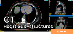
OARs** (Included in Thorax/Breast)
- Ascending Aorta (Thorax)
- Atria
- Coronary Sinus ventricles
- Main coronary artery left
- Vena cava superior
- Ventricles
- Ventricle left sections
- Anterior
- Apical
- Inferior
- Lateral
- Septal
**Guideline: Duane et al (2017), Lee et al (2017)
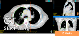
OARs**
- Brachial plexus
- Bronchial tree
- Carina
- Bronchia
- Bronchus
- Chest walls
- Esophagus
- Heart
- LAD artery (left anterior descending)
- Liver
- Pericardium
- Pulmonary arteries
- Skin
- Spinal cord
- Trachea
- Vena cava
**Guideline: UK SABR Consortium (2019), De Rose et al, (2017)
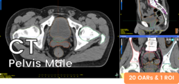
OARs*
ROI**
- Anal canal
- Bladder
- Bowel bag
- Duodenum
- Femoral head (L/R)
- Iliac (L/R)
- Kidney (L/R)
- Liver
- Large bowel
- Medullar canal
- Penile bulb
- Prostate
- Rectum
- Seminal vesicle
- Sigmoid
- Small bowel
- Spinal cord
- Stomach
- CTVn prostate
*Guideline: Gay et al (2012)
**Region of interest

OARs*
LNs & ROI**
- Anal Canal
- Bladder
- Bowel bag
- Femoral head (L/R)
- Iliac (L/R)
- Kidney (L/R)
- Large bowel
- Liver
- Medullar canal
- Parametrium
- Rectum
- Sigmoid
- Small bowel
- Spinal cord
- Stomach
- Vagina
- Common Iliac Gyneco LNs
- CTVt Gyneco
- Iliac Gyneco LN (L/R)
- Inguinal gyneco LN (L/R)
- Lomboartic gyneco LNs
- Presacral gyneco LNs
*Guideline: Gay et al (2012)
**Region of interest
Want to discover more?
Be Automated
Automate all steps of the segmentation process with our Batch mode. Have the automatic contouring done in the background and exported directly from the scanner to the TPS of your choice.
Be Thorough
Have more than 200 OARs and exclusive models for CTV including lymph nodes automatically delineated at the power of AI. No structures or slices forgotten. All you have to do is to review and validate.
Be Faster
Obtain highly accurate full-body delineation in 3 minutes. Save up to 90% of your contouring time. Reduce the overload of your clinic staff to focus what matters most: your patients.